Did you know that an MRI of the brain can unveil hidden insights about your cognitive health? As our understanding of brain wellness grows, so does the significance of advanced imaging techniques like MRI. Whether you’re experiencing unexplained headaches, cognitive decline, or neurological symptoms, a doctor may recommend this non-invasive procedure to explore potential underlying conditions. Understanding why these scans are ordered can empower you in discussions with your healthcare provider and help you manage concerns about your brain health. In this article, we will delve into the medical reasons for an MRI of the brain, illuminating how this valuable diagnostic tool can guide treatment and enhance your well-being.
Understanding the Importance of Brain MRIs

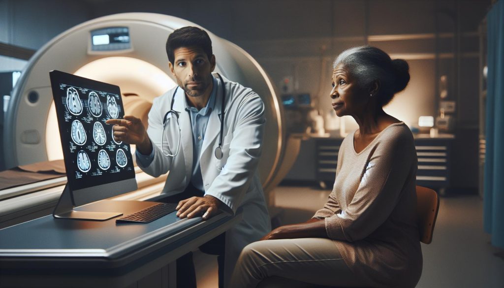
Brain MRI scans stand as a pivotal tool in modern medicine, providing unparalleled insights into the intricate workings of our brains. With their exceptional ability to produce detailed images, these scans allow healthcare professionals to visualize not just the physical structures of the brain but also to investigate a myriad of medical conditions that can affect cognitive functions and overall neurological health. This non-invasive procedure is crucial for early detection and accurate diagnosis, making it an indispensable part of neurological assessments.
When a doctor orders a brain MRI, it often stems from a range of symptoms or concerns that could indicate a serious underlying issue. Some of the most common medical reasons include persistent headaches, unexplained cognitive issues, or neurological symptoms like weakness, numbness, or vision problems. By capturing detailed images of brain tissues, MRIs facilitate the identification of various conditions such as tumors, strokes, and neurodegenerative diseases, thus laying the groundwork for timely and effective treatment.
Understanding the role of brain MRIs in evaluating conditions is essential for anyone looking to prioritize their brain health. While it’s normal to feel apprehensive about the procedure, knowing its importance can transform anxiety into empowerment. Recognizing the signs that warrant an MRI, such as severe headaches or sudden changes in cognitive function, can hasten your journey to optimal health. Brain MRIs not only illuminate potential problems but also aid in crafting tailored treatment plans, thereby enhancing patient outcomes and supporting cognitive wellness.
Common Medical Reasons for Brain MRIs
When it comes to assessing brain health, a magnetic resonance imaging (MRI) scan can be a crucial diagnostic tool. From persistent headaches to unexplained cognitive shifts, there are numerous reasons why a healthcare provider may recommend a brain MRI. Each of these scenarios sheds light on the significant role that MRIs play in uncovering potential medical issues and guiding effective treatments.
Among the most common reasons for a brain MRI is the investigation of persistent headaches. While occasional headaches may not be cause for concern, chronic or severe headaches can signal underlying conditions such as migraines, tumors, or increased intracranial pressure. By creating detailed images of the brain, MRIs help identify structural abnormalities, enabling physicians to pinpoint the cause and recommend appropriate treatments.
Another medical scenario that frequently prompts an MRI involves neurological symptoms such as weakness, numbness, or changes in vision. These symptoms can stem from a variety of issues, including stroke, multiple sclerosis, or nerve damage. In these cases, MRIs serve as a non-invasive method to visualize the brain and spinal cord, allowing healthcare providers to assess the extent of any damage and devise a comprehensive management plan.
Additionally, cognitive decline or sudden changes in mental status are critical indicators that may necessitate a brain MRI. Symptoms such as confusion, memory loss, or difficulty concentrating could suggest neurodegenerative diseases like Alzheimer’s or other forms of dementia. Timely imaging allows for early diagnosis, enabling interventions that can significantly enhance the quality of life and cognitive health.
In summary, a brain MRI can provide invaluable insights into a multitude of medical scenarios, from headache evaluations to neurological assessments. By understanding the reasons behind an MRI referral, patients can feel more empowered about their health and make informed decisions regarding their care. Remember, if you experience any concerning symptoms, it’s crucial to consult a qualified healthcare professional who can guide you through the necessary steps for evaluation and treatment.
How MRIs Help Diagnosing Neurological Conditions

Understanding the intricacies of brain health is crucial, especially considering that neurological conditions can often present with subtle symptoms that may escalate into serious issues if left unchecked. Magnetic resonance imaging (MRI) plays a pivotal role in diagnosing a range of neurological disorders, acting as a powerful tool that captures detailed images of the brain’s structure. This imaging technique allows healthcare providers to detect abnormalities that might signal underlying conditions, fostering timely intervention.
One of the most significant advantages of MRI in neurology is its non-invasive nature coupled with its high-resolution imaging capabilities. This means that doctors can visualize the brain without needing surgical procedures, allowing them to explore potential causes of symptoms such as chronic headaches, blurred vision, or sudden changes in mental acuity. For instance, in cases of suspected multiple sclerosis, MRI can reveal lesions on the brain and spinal cord, which are critical for confirming a diagnosis. Identifying these features is essential for implementing effective management strategies early in the disease process.
An MRI can also aid in differentiating between various types of neurological disorders. Symptoms like numbness or weakness might suggest issues ranging from stroke to peripheral nerve damage. With the help of contrast-enhanced MRI scans, conditions such as tumors and infections can be identified much faster, leading to quicker treatment decisions. Moreover, the use of functional MRI (fMRI) allows for assessment of brain activity by measuring changes in blood flow, providing insights into functional deficits that might correlate with clinical symptoms. This level of detail is invaluable for creating tailored treatment plans that address specific patient needs.
In the realm of workplace wellness, understanding these diagnostic tools opens up discussions on how we can better care for our cognitive health. For instance, fostering an environment that encourages breaks, mental health awareness, and ergonomic setups can help alleviate some symptoms that lead to MRI referrals in the first place. Incorporating healthy habits into your daily routine-like regular exercise, a balanced diet, and stress management techniques-can significantly contribute to maintaining brain health and potentially reducing the need for diagnostic imaging. Always remember, however, that if any concerning symptoms arise, seeking professional medical advice is the best course of action.
The Role of MRI in Evaluating Head Injuries
Head injuries can have lasting effects on cognitive function, emotional health, and overall well-being. MRI is an essential tool in evaluating the extent of brain damage as it provides detailed images of the brain’s structure, allowing healthcare professionals to identify possible injuries like concussions, contusions, or more severe traumatic brain injuries (TBIs). This non-invasive imaging method excels at detecting subtle changes that can occur in brain tissues and structures, making it invaluable for diagnosing conditions that may not be immediately apparent.
One of the significant benefits of MRI in head injury assessments is its ability to highlight areas of hemorrhage or bleeding, which could necessitate immediate medical intervention. For example, following a car accident or a fall, a patient may report symptoms such as headaches, confusion, or dizziness. An MRI can visualize a hematoma, which could indicate significant bleeding that requires surgery. Additionally, MRI scans can reveal injuries to the brain’s white matter. This is particularly important as damage in these areas can lead to cognitive deficits that might not surface until later.
Patients often wonder when an MRI is necessary after a head injury. Generally, doctors will recommend this imaging study if there are signs of serious complications-such as prolonged unconsciousness, severe headaches not responding to medication, seizure activity, or any neurological deficits. It is also a crucial tool during follow-up evaluations to monitor recovery progress over time. For instance, regular MRI assessments can help identify whether a patient is healing as expected or if new issues are developing, thus guiding further treatment or rehabilitation strategies.
Understanding how to care for your brain in the context of workplace wellness can facilitate recovery and minimize the risk of future injuries. Prioritizing a safe working environment, such as using ergonomic furniture, can prevent falls that may lead to head injuries. Encouraging practices like regular breaks and stress management exercises can also promote cognitive health, helping individuals be more aware of their mental well-being. Remember, while many head injuries may resolve with time, seeking medical advice and undergoing appropriate imaging when necessary is crucial to ensure comprehensive care.
When to Consider MRI for Migraines and Headaches
Migraines can be debilitating, impacting daily activities and overall quality of life. When persistent headaches fail to respond to standard treatments or are accompanied by unusual symptoms, it may be time to consider more in-depth evaluations, including an MRI of the brain. An MRI can help identify potential underlying issues that may be contributing to these headaches beyond the usual migraine triggers.
It is particularly important to discuss MRI imaging with your doctor when headaches come with neurological symptoms such as vision changes, difficulty speaking, weakness in limbs, or confusion. These could indicate serious conditions like strokes, tumors, or other structural abnormalities in the brain that require immediate attention. Additionally, if headaches are significantly more severe than typical migraine episodes or have a sudden onset, an MRI can provide vital information to rule out any serious conditions, ensuring timely treatment.
Furthermore, MRI can be useful to distinguish between different types of headaches, such as tension-type headaches versus migraines or other more complex headache disorders like cluster headaches. By visualizing the brain’s anatomy in high detail, MRIs can help healthcare professionals tailor better treatment plans that target the specific causes of an individual’s headache condition. This approach can be particularly empowering for patients seeking to understand their health and pursue appropriate interventions.
Incorporating lifestyle adjustments alongside medical evaluation is essential. Maintaining a consistent routine, addressing stress through mindfulness practices, and ensuring ergonomic setups in the workplace can mitigate migraine triggers. These strategies not only improve overall well-being but also promote cognitive health, enhancing your ability to manage headaches effectively. Always consult healthcare professionals for advice on imaging and treatments tailored to specific health needs.
MRI as a Tool for Detecting Brain Tumors
Detecting brain tumors early can significantly improve treatment outcomes and survival rates. Magnetic Resonance Imaging (MRI) stands out as one of the most effective tools in the medical imaging arsenal, providing detailed images of the brain’s structure while allowing physicians to observe abnormalities with precision. Tumors may manifest through various symptoms, such as persistent headaches, seizures, or changes in vision or cognitive function, prompting a doctor to order an MRI. High-resolution images enable doctors to identify the presence of tumors, their size, and even their location within the brain, laying the groundwork for a targeted treatment plan.
Innovations in MRI technology, such as functional MRI (fMRI) and diffusion tensor imaging (DTI), enhance its utility in detecting brain tumors. While traditional MRI assesses the structural aspects of the brain, fMRI measures brain activity by detecting changes in blood flow, which may help in understanding how a tumor affects specific brain functions. DTI allows for the visualization of white matter tracts, aiding in the assessment of how a tumor may impact connections between different brain regions. This layered approach equips healthcare providers with a deeper understanding of tumor behavior and potential impact on neurological function.
H3: Importance of Timely Detection
Early detection of brain tumors is crucial for devising an effective treatment strategy. An MRI can guide surgical plans, radiation therapy, or other interventions. Furthermore, discussing MRI results with healthcare professionals not only facilitates swift decision-making but also helps patients understand the tumor’s nature-whether benign or malignant-and the best pathways for management. Being informed empowers patients, enabling them to participate actively in their health decisions, which can alleviate stress and provide a clearer roadmap to recovery.
To foster cognitive health alongside medical interventions, consider adopting supportive lifestyle changes. Simple practices like regular physical activity, mindfulness meditation, and a balanced diet rich in antioxidants can enhance overall brain health. These strategies not only help mitigate stress-integral when navigating health challenges-but also boost cognitive resilience, thereby promoting a holistic approach to well-being. Always prioritize thorough discussions with healthcare professionals when exploring options related to imaging, treatment, or lifestyle adjustments, ensuring that each decision contributes to your health goals.
Interpreting MRI Results: What to Expect
Understanding your MRI results can be pivotal in addressing health concerns, particularly those affecting the brain. When your doctor orders an MRI, it’s generally due to specific symptoms or conditions that warrant detailed imaging, such as persistent headaches, seizures, or cognitive changes. Once the MRI is complete, the next step involves interpreting these complex images, often underscored by the anxiety of waiting for results. It’s crucial to remember that MRI scans provide high-resolution images that help radiologists and neurologists identify structural abnormalities within the brain, contributing to a clearer diagnostic picture.
Radiologists analyze the MRI images for various indicators, such as the presence of tumors, signs of stroke, swelling, or structural anomalies like lesions. They may categorize findings by their appearance and context-whether they suggest a benign condition, a malignancy, or other neurological issues. Following the analysis, detailed reports are generated, often using non-technical language where possible, to ensure that you can understand your medical condition better. It’s essential to engage with your healthcare professional during this process, asking questions to clarify any parts of the report that concern or confuse you.
Moreover, while interpreting results, consider that not all findings may require immediate action. For example, certain structural variations in the brain can be normal and not indicative of a disease. Understanding the context is vital; what is significant in one patient’s circumstances might not be in another’s. The more informed you are about what the results mean, the more empowered you can be in making decisions about your care. This proactive approach reduces anxiety and promotes a productive dialogue with your healthcare team.
As you navigate this journey, remember to prioritize your mental well-being. Stress management techniques-such as mindfulness, regular physical activity, and open communication with loved ones-can significantly boost your cognitive resilience during challenging times. Engaging in discussions with professionals about lifestyle changes can also be beneficial for your overall brain health. Embrace this time as an opportunity to foster a deeper understanding of your body and take charge of your health journey.
Innovations in MRI Technology for Brain Imaging
The field of MRI technology is advancing rapidly, significantly enhancing our ability to diagnose and treat brain-related conditions. One of the most exciting innovations in recent years is the development of functional MRI (fMRI), which goes beyond standard imaging to capture changes in brain activity. This technology measures blood flow changes related to neural activity, enabling clinicians to observe brain function in real-time. For instance, fMRI can help localize the regions of the brain responsible for critical functions such as movement, speech, and memory, which is invaluable for surgical planning or rehabilitation strategies post-injury.
Another groundbreaking advancement is the introduction of ultra-high-field MRI scanners. These devices, which operate at higher magnetic field strengths (such as 7 Tesla), provide unprecedented image resolution and contrast, allowing for more precise identification of small tumors, minute vascular changes, or subtle neurological disorders. For example, they can reveal fine anatomical details that traditional 1.5 or 3 Tesla machines might miss, giving radiologists greater accuracy in diagnosing conditions like early-stage multiple sclerosis or detecting abnormalities following trauma.
Moreover, developments in software algorithms have also improved image reconstruction techniques, reducing scan times while enhancing image quality. As a result, patients experience less time in the scanner, which is especially beneficial for those who may have difficulty staying still or experience claustrophobia. These advancements not only ease patient anxiety but also increase the throughput of imaging departments, allowing more individuals to access these essential diagnostic tools.
Lastly, innovations such as artificial intelligence (AI) are beginning to play a significant role in MRI interpretation. AI algorithms can process large datasets quickly, assisting radiologists in identifying patterns and anomalies that might otherwise be overlooked. This not only improves diagnostic accuracy but can also streamline workflow in busy imaging centers, allowing healthcare professionals to focus on patient care rather than administrative tasks.
As these technologies continue to evolve, they hold the promise of not only enhancing diagnostic capabilities but also allowing for more personalized and effective treatment pathways for various neurological conditions. Staying informed about these advancements can empower patients and healthcare advocates to take an active role in discussions with their providers, ensuring they receive the best care possible.
Preparing for Your Brain MRI Appointment
Preparing for a brain MRI can feel daunting, but understanding what to expect and how to prepare can significantly reduce anxiety. Brain MRIs are crucial tools for diagnosing a variety of neurological conditions, whether you’re experiencing symptoms like headaches, dizziness, or unresolved cognitive issues. Knowing that this non-invasive procedure uses powerful magnets and radio waves to create detailed images of your brain can help you feel more at ease as you approach your appointment.
Before your MRI, it’s essential to adhere to certain guidelines to ensure the process runs smoothly. Here are some key preparation steps:
- Consult Your Doctor: Discuss any medications you’re taking, allergies, or health conditions with your doctor. They may have specific recommendations based on your situation.
- Dress Comfortably: Wear loose-fitting clothing without metal fasteners. Many facilities provide gowns for patients to wear during the scan.
- Remove Metal Objects: Ensure to remove all jewelry, glasses, and any metal implants or devices that might interfere with the MRI.
- Discuss Claustrophobia: If you experience anxiety in enclosed spaces, inform your medical team beforehand. They may provide relaxation techniques or sedation options.
- Plan for the Duration: MRIs typically last between 20 to 60 minutes. Bring something to help you stay occupied afterward, as you might need to wait for your results.
Finally, approaching the MRI with a positive mindset can be beneficial. Techniques such as deep breathing, visualization, or listening to calming music (if permitted) during the scan may help reduce stress levels. Remember to communicate any concerns with your healthcare provider-your comfort is paramount. Proper preparation can transform the experience from a source of stress into a straightforward step toward understanding and improving your brain health.
Safety and Risks Associated with Brain MRIs
While brain MRIs are generally considered safe and effective for diagnosing various neurological conditions, it is important to be aware of potential safety concerns and risks associated with this procedure. The non-invasive nature of MRI technology, which employs strong magnetic fields and radio waves to create detailed images, significantly reduces the likelihood of complications compared to invasive imaging methods. However, some factors should be considered to ensure a safe experience.
One of the primary concerns involves the presence of metal in or on the body, as the strong magnets used in MRIs can interfere with metallic implants and devices. Patients with certain implants, such as pacemakers, cochlear implants, or other electronic or metal components, must disclose this information to their healthcare provider prior to the procedure. Alternatives or additional precautions may need to be taken for individuals in these situations. Additionally, individuals should be cautious about wearing clothing with metal fasteners or carrying metal objects into the MRI room.
Another concern is for those who may experience anxiety or claustrophobia during the scan. The narrow tube of the MRI machine can feel confining, and some individuals may feel uncomfortable or distressed. It’s wise to discuss any fears with the medical personnel before the appointment, who can provide options for calming techniques, music, or even sedation if necessary.
Lastly, while MRI scans do not involve radiation exposure like CT scans, some patients may still experience mild side effects such as temporary discomfort from lying still for a prolonged period or a sensation of warmth due to the magnetic field. If you have any concerns about these potential risks or your individual health circumstances, it is always best to consult with your healthcare provider to ensure that brain MRI is the right and safest option for your situation.
Alternatives to MRI for Brain Evaluation
In some cases, a brain MRI may not be the only or best choice for evaluating brain health, and alternative imaging techniques can provide valuable insights. Each method has its strengths and limitations, depending on the condition being assessed, the patient’s health profile, and specific diagnostic needs.
CT Scans
Computed Tomography (CT) scans use X-rays to create cross-sectional images of the brain. This method is particularly useful for quickly diagnosing conditions like strokes, bleeding, and fractures. CT scans are often preferred in emergency settings due to their speed, making them ideal for assessing acute head injuries or sudden neurological symptoms. However, they expose patients to a small amount of radiation, which is a significant consideration.
Ultrasound
For certain populations, such as infants, brain ultrasound can be an effective alternative. This technique uses sound waves to produce images of the brain and is especially useful for detecting structural abnormalities in newborns, thanks to their fontanelles (soft spots in the skull). It is non-invasive and does not involve radiation, making it a safe option for infants.
Functional MRI (fMRI)
Functional MRIs provide a different perspective by measuring brain activity through changes in blood flow. Although fMRI is not typically used as a primary diagnostic tool, it has significant applications in research and pre-surgical planning. It helps understand brain function and delineates areas responsible for critical tasks such as movement or language, providing essential insights, especially before surgery.
Electroencephalogram (EEG)
An EEG records electrical activity in the brain and can be instrumental in diagnosing epilepsy, sleep disorders, and monitoring brain function during surgery. While it doesn’t provide anatomical images, it offers crucial information about brain activity patterns, which may indicate underlying conditions that require further evaluation.
In summary, while MRI is an invaluable tool for brain evaluation, alternatives such as CT scans, ultrasounds, fMRI, and EEGs can complement or provide necessary insights for specific conditions. Always consult with healthcare providers to determine the most appropriate imaging method based on individual health needs and circumstances.
Post-MRI Follow-Up: Next Steps After Your Scan
After undergoing a brain MRI, it’s crucial to understand what comes next as you await your results and navigate the potential implications for your health. The MRI scans provide detailed images that can reveal issues such as tumors, inflammation, or structural anomalies, which can help healthcare providers make informed decisions about your care. While the anticipation may feel overwhelming, taking proactive steps can empower you during this period.
First, ensure that you follow up with your healthcare provider as soon as possible to discuss your results. Scheduling this appointment can help you gain clarity on any findings, and your doctor can guide you on the next steps based on your specific situation. Be prepared to ask questions regarding any terms or conditions you might not understand; having a clear dialogue is essential for comprehensive care.
Tips for Managing Your Well-Being
While waiting for your results, it’s important to manage stress and maintain a healthy mindset. Here are some practical strategies to consider:
- Mindfulness and Relaxation: Engaging in mindfulness exercises, such as meditation or deep-breathing techniques, can help reduce anxiety. Setting aside a few minutes each day for relaxation can enhance your mental clarity and overall well-being.
- Physical Activity: Regular physical activity has been proven to boost mood and cognitive function. Consider incorporating activities such as walking, yoga, or even light stretching to help alleviate stress.
- Stay Informed but Balanced: While it’s natural to research your symptoms or potential diagnoses, be cautious about information overload. Seek reputable sources and avoid falling into the trap of excessive online searches that may lead to unnecessary anxiety.
What If Further Tests Are Needed?
Sometimes, additional imaging or diagnostic tests may be necessary following your MRI. Your doctor may recommend further assessments such as a CT scan, fMRI, or even biopsies to gather more information about your condition. Understanding the reasoning behind these recommendations can help alleviate concerns, as they are often essential steps to ensure accurate diagnosis and targeted treatment.
This time of uncertainty can be challenging, but remember that you are not alone. Lean on your support network of family and friends, and consider communicating your feelings about the waiting period. Your proactive approach in seeking information and remaining engaged with your healthcare can play a significant role in navigating your health journey effectively.
FAQ
Q: What symptoms might lead a doctor to order a brain MRI?
A: Doctors may order a brain MRI if a patient exhibits symptoms like persistent headaches, sudden seizures, unexplained dizziness, or changes in vision. These symptoms can indicate underlying neurological issues that need further evaluation.
Q: How does an MRI help in diagnosing multiple sclerosis?
A: An MRI is essential for diagnosing multiple sclerosis (MS) as it can reveal lesions or plaques in the brain and spinal cord. These images assist doctors in confirming the presence and extent of MS, guiding treatment options.
Q: Can an MRI detect mental health disorders?
A: While MRIs cannot diagnose mental health disorders directly, they can help rule out structural brain issues that may contribute to conditions like depression or anxiety. Neurologists may use MRI results to understand better the underlying brain function.
Q: Why might an MRI be preferred over a CT scan for brain evaluation?
A: MRI is often preferred over CT scans for brain evaluation due to its superior ability to visualize soft tissues and detect abnormalities without radiation exposure. This makes MRIs safer for repeated use, especially in ongoing neurological assessments.
Q: What role does MRI play in assessing brain tumors?
A: MRI is crucial in assessing brain tumors as it provides detailed images that help identify tumor size, location, and type. This information is vital for planning treatment strategies, including surgery or radiation therapy.
Q: Is MRI useful in evaluating the effects of a stroke?
A: Yes, MRI is instrumental in evaluating stroke effects. It helps identify area(s) of the brain affected by the stroke, determines its type (ischemic or hemorrhagic), and guides further treatment decisions.
Q: How should one prepare for a brain MRI appointment?
A: Preparation for a brain MRI typically includes wearing comfortable clothing without metal, informing your doctor of any implants or relevant health conditions, and possibly fasting for a few hours before the procedure, based on the clinic’s guidelines.
Q: What are the risks associated with brain MRIs?
A: While MRIs are generally safe, risks include potential discomfort from the closed space of the machine and rare allergic reactions to contrast agents. It’s essential to discuss any concerns with your healthcare provider prior to the scan.
Closing Remarks
Understanding the reasons behind a doctor’s decision to order an MRI of the brain can empower you to take proactive steps in monitoring your brain health. Whether it’s to investigate unexplained symptoms or rule out serious conditions, an MRI plays a crucial role in medical diagnostics. If you have lingering questions or concerns about brain health and the significance of imaging, don’t hesitate to explore our detailed guides on workplace wellness and cognitive performance.
Ready to prioritize your brain health? Consider signing up for our newsletter to receive expert tips on stress management, ergonomic workspace setups, and biohacking techniques that promote cognitive well-being. Your health is worth it! For further resources, check out our articles on neuroplasticity and effective stress management strategies or explore additional imaging options related to your health concerns. Engage with us-share your thoughts in the comments below, and join our community dedicated to fostering better brain health together.